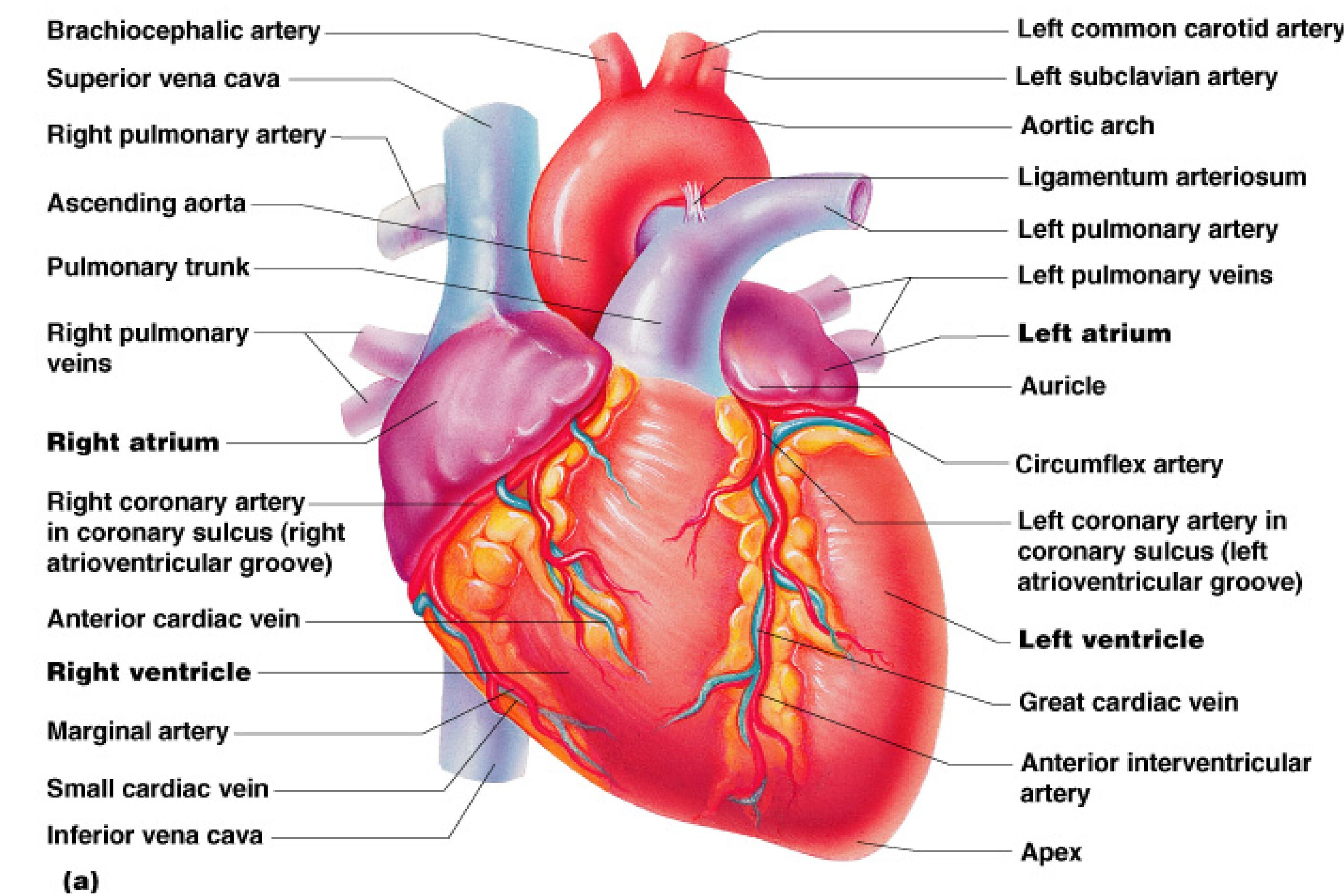
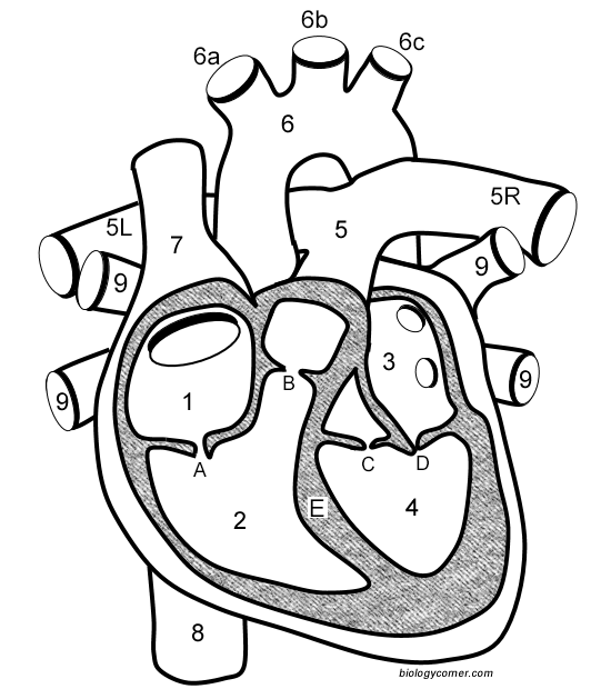
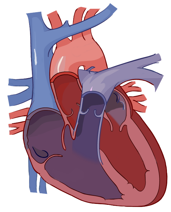
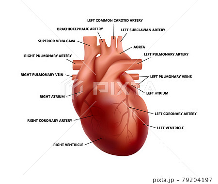
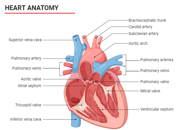
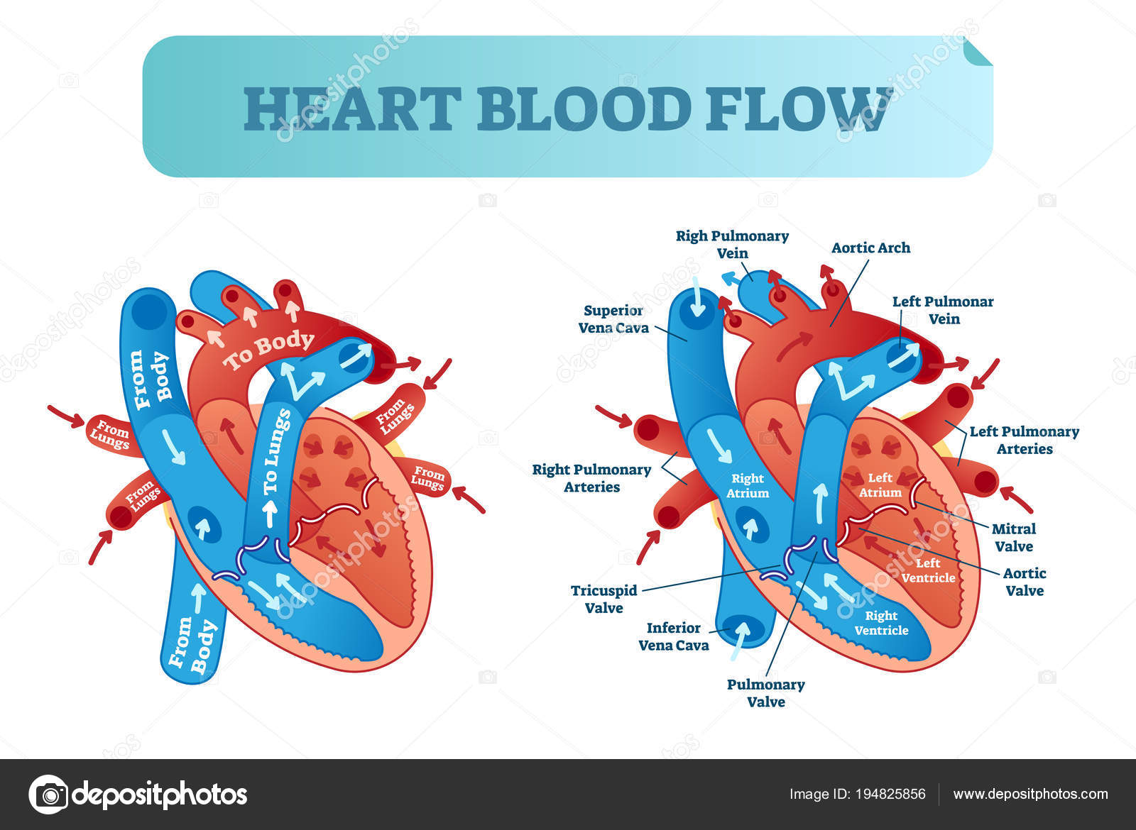
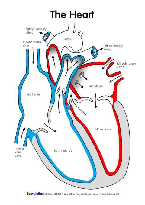
45 heart diagram labeled
Label the heart — Science Learning Hub In this interactive, you can label parts of the human heart. Drag and drop the text labels onto the boxes next to the diagram. Selecting or hovering over a box will highlight each area in the diagram. pulmonary vein semilunar valve right ventricle right atrium vena cava left atrium pulmonary artery aorta left ventricle Download Exercise Tweet Diagram of Blood Flow Through the Heart - Bodytomy If not, you can have a look at the labeled diagram of the human heart present in this article. The strongest muscle in the human body is the human heart. The human heart continues to pumps liters of blood throughout the body all lifelong. The minute the heart stops pumping blood, it leads to a heart attack that can prove to be fatal. Blood Circulation within the Heart for Kids. …
› Heart-Diagram-LabeledDiagram of Human Heart and Blood Circulation in It | New ... Nov 27, 2022 · The outermost layer of your heart wall is called the epicardium, which is basically a very thin layer of serous membrane. The membrane provides lubrication and protection to the outer side of your heart, as you can see in heart diagram labeled. Myocardium. Right beneath epicardium is another relatively thicker layer called myocardium.
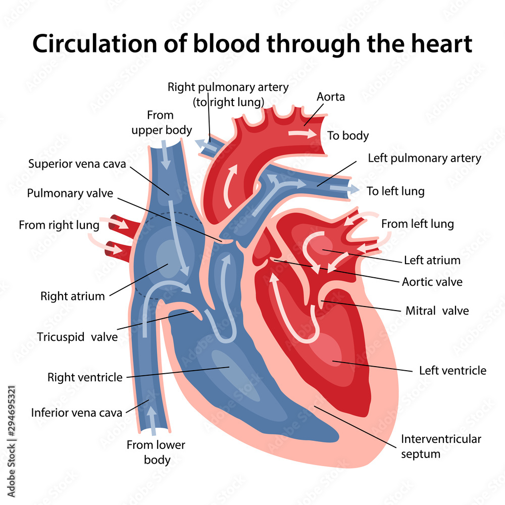
Heart diagram labeled
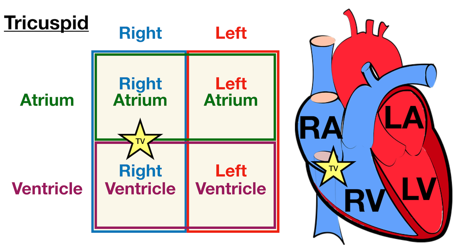
Heart diagram labeled Flashcards | Quizlet Only $35.99/year Heart diagram labeled Flashcards Learn Test Match Flashcards Learn Test Match Created by Mike_Longo2 Terms in this set (13) 1. Superior Vena Cava deoxygenated the body 2. Pulmonary Artery - - deoxygenated blood to the lungs 3. Pulmonary Vein oxygenated blood from the lungs 4. Mitral Valve 4. Mitral Valve 5. Aortic Valve 5. Heart Blood Flow | Simple Anatomy Diagram, Cardiac Circulation ... - EZmed Diagram: Blood flow through the right side of the heart involving the following cardiac structures: superior vena cava (SVC), inferior vena cava (IVC), right atrium (RA), tricuspid valve (TV), right ventricle (RV), pulmonary valve (PV), and main pulmonary artery (PA). Trick to Remember the Right Side › blog › heart-anatomy-labeledHeart Anatomy: Labeled Diagram, Structures, Blood Flow ... There are 4 chambers, labeled 1-4 on the diagram below. To help simplify things, we can convert the heart into a square. We will then divide that square into 4 different boxes which will represent the 4 chambers of the heart. The boxes are numbered to correlate with the labeled chambers on the cartoon diagram. View fullsize
Heart diagram labeled. The Heart | Circulatory Anatomy - Visible Body 1. The Heart Wall Is Composed of Three Layers. The muscular wall of the heart has three layers. The outermost layer is the epicardium (or visceral pericardium). The epicardium covers the heart, wraps around the roots of the great blood vessels, and adheres the heart wall to a protective sac. The middle layer is the myocardium. Human Heart Diagram - Side View and Top View - Heart Valve Surgery Here is a human heart diagram showing a top view of the heart - as if you were looking down on the heart. As shown below, this human heart diagram clearly illustrates the valves of the heart. The valves illustrated below are the pulmonary, tricuspid, aortic and mitral valve. So you know, I had the aortic and pulmonary valves of my heart ... 15,349 Human Heart Diagram Images, Stock Photos & Vectors - Shutterstock Find Human heart diagram stock images in HD and millions of other royalty-free stock photos, illustrations and vectors in the Shutterstock collection. Thousands of new, high-quality pictures added every day. Human Heart - Diagram and Anatomy of the Heart - Innerbody The heart wall is made of 3 layers: epicardium, myocardium and endocardium. Epicardium. The epicardium is the outermost layer of the heart wall and is just another name for the visceral layer of the pericardium. Thus, the epicardium is a thin layer of serous membrane that helps to lubricate and protect the outside of the heart.
anatomylearner.com › goat-anatomyGoat Anatomy – External and Internal ... - AnatomyLearner Jul 21, 2021 · Goat Anatomy – External and Internal Anatomical Features with Labeled Diagram 03/11/2022 21/07/2021 by anatomylearner Veterinary students and farm owners might know the external and internal goat anatomy to maintain good health and increase the productivity of a herd. Circulatory System Diagram - Cardiovascular System and Blood ... Circulatory system diagrams are visual representations of the circulatory system, also referred to as the cardiovascular system. It is comprised of three parts: the pulmonary circulation, coronary circulation, and systemic circulation. The main function of the circulatory system is to circulate blood, which carries oxygen and nutrients ... Heart Labeling Quiz: How Much You Know About Heart Labeling? Start. Create your own Quiz. Here is a Heart labeling quiz for you. The human heart is a vital organ for every human. The more healthy your heart is, the longer the chances you have of surviving, so you better take care of it. Take the following quiz to know how much you know about your heart. Heart diagram labelling quiz - ESL Games Plus Heart diagram labelling quiz When you make a closed fist, it will roughly be the size of your own heart. In addition, if you close and open your fists around 60 times per minute - the normal resting heart rate for adults - you can catch a glimpse at how hard the heart works to pump blood throughout your body. The heart is an extremely vital organ.
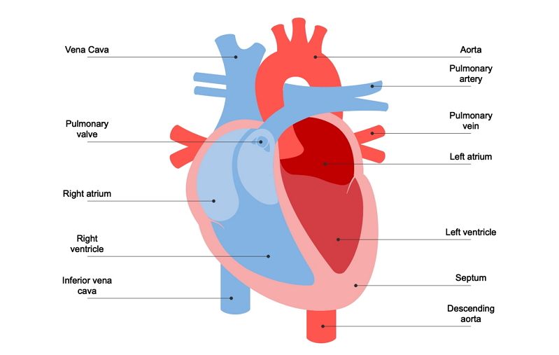
Heart Diagram with Labels and Detailed Explanation - BYJUS Well-Labelled Diagram of Heart The heart is made up of four chambers: The upper two chambers of the heart are called auricles. The lower two chambers of the heart are called ventricles. The heart wall is made up of three layers: The outer layer of the heart wall is called epicardium. The middle layer of the heart wall is called myocardium. Male Human Anatomy Diagram Pictures, Images and Stock Photos Pacemaker Diagram Cross section of a human heart with pacemaker fitted, showing the major arteries and veins. This is an EPS 10 vector illustration and includes a high resolution JPEG. male human anatomy diagram stock illustrations . Pacemaker Diagram. Cross section of a human heart with pacemaker fitted, showing the major arteries and veins. This is an EPS 10 vector … 13+ Heart Diagram Templates - Sample, Example, Format Download Color Heart Diagram Sample Format Free Download cdhb.health.nz This colored heart diagram is a graphic representation of the organ which can be used for presentations and videos about the subject of human heart. The picture is in a coloured format and is available for a free download. Free Download Inner Part Of The Heart Sample Diagram Human Heart (Anatomy): Diagram, Function, Chambers, Location in Body The heart has four chambers: The right atrium receives blood from the veins and pumps it to the right ventricle. The right ventricle receives blood from the right atrium and pumps it to the...
Unlabelled heart diagram - Healthiack Matej G. is a health blogger focusing on health, beauty, lifestyle and fitness topics. He has been with healthiack.com since 2012 and has written and reviewed well over 500 coherent articles.
Heart | Structure, Function, Diagram, Anatomy, & Facts In humans and other mammals and in birds, the heart is a four-chambered double pump that is the centre of the circulatory system. In humans it is situated between the two lungs and slightly to the left of centre, behind the breastbone; it rests on the diaphragm, the muscular partition between the chest and the abdominal cavity. Britannica Quiz
A Diagram of the Heart and Its Functioning Explained in Detail The heart blood flow diagram (flowchart) given below will help you to understand the pathway of blood through the heart.Initial five points denotes impure or deoxygenated blood and the last five points denotes pure or oxygenated blood. I hope, the heart diagram and the blood flow chart given above is clear to you.
A Labeled Diagram of the Plant Cell and Functions of its Organelles You can save and print this diagram of the plant cell. It will help you with your revision. That’s about how the organelles in a cell function. It’s unbelievable how a tiny cell can help a full-grown plant to grow and produce energy. As they say, life originates in a single cell and it is this cell that plays an important role in the growth ...
› nflNFL News, Scores, Standings & Stats | FOX Sports Get NFL news, scores, stats, standings & more for your favorite teams and players -- plus watch highlights and live games! All on FoxSports.com.
biologywise.com › labeled-plant-cell-diagram-functionsA Labeled Diagram of the Plant Cell and Functions of its ... Known to be the heart of the cell, the nucleolus transcribes ribosomal RNA. It is composed of proteins and nucleic acid and is known to be a genetically determined element. Function: Produces ribosomes. Peroxisomes. Membrane-bound packets of oxidative enzymes, the peroxisomes play a vital role in converting fatty acids to sugar.
A Diagram of the Heart and Its Functioning Explained in Detail In this article, we are going to discuss various functions of heart with the help of a well labeled diagram. This diagram of the heart will not only give you details about the various parts, but will also explain the importance of keeping your heart healthy. Human heart is slightly bigger than the size of one’s fist. It is situated at a very ...
Heart Anatomy: Labeled Diagram, Structures, Blood Flow 24.02.2021 · Function and anatomy of the heart made easy using labeled diagrams of cardiac structures and blood flow through the atria, ventricles, valves, aorta, pulmonary arteries veins, superior inferior vena cava, and chambers. Includes an exercise, review worksheet, quiz, and model drawing of an anterior vi
› male-human-anatomy-diagramMale Human Anatomy Diagram Pictures, Images and Stock Photos Labeled Anatomy Chart of Shoulder, Elbow and Triceps Muscles in Skeleton on Black Background Labeled human anatomy diagram of man's shoulder bones, triceps muscles and connective tissue in a posterior view on a black background. male human anatomy diagram stock pictures, royalty-free photos & images
Diagram of Human Heart and Blood Circulation in It 27.11.2022 · The function of heart is quite complex, but you can understand things better through the heart diagram labeled below. It provides information about different chambers of the heart and valves that help transfer blood from one part of your heart to another. Keep reading to learn more about how your heart works. Four Chambers of the Heart and Blood Circulation. The …
Conduction System of the Heart: Step-By-Step Labeled Diagram … 05.10.2020 · Easily learn the conduction system of the heart using this step-by-step labeled diagram. The cardiac conduction system is the electrical pathway of the heart that includes, in order, the SA node, AV node, bundle of His, bundle branches, and Purkinje fibers. Learn about pacemaker cells and cardiac action potentials causing contraction and ...
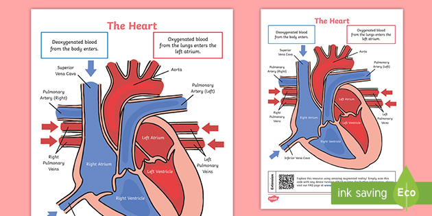
Labelling the heart — Science Learning Hub Labelling the heart. The heart is a muscular organ that pumps blood through the blood vessels of the circulatory system. Blood transports oxygen and nutrients to the body. It is also involved in the removal of metabolic wastes. In this activity, students use online and paper resources to identify and label the main parts of the heart.
A Labeled Diagram of the Human Heart You Really Need to See A Labeled Diagram of the Human Heart You Really Need to See. The heart, one of the most significant organs in the human body, is nothing but a muscular pump which pumps blood throughout the body. The human heart and its functions are truly fascinating. The heart, though small in size, performs highly significant functions that sustains human life.
Label the HEART | Circulatory System Quiz - Quizizz True or False: Blood flows in the following sequence in the heart: Vena cava, right atrium, right ventricle, pulmonary artery, lungs, pulmonary veins, left atrium, left ventricle, aorta. Q. True or False: There are four chambers in the heart. Q. Place the pathway of blood through the heart in the correct sequence. Q.
The Anatomy of the Heart, Its Structures, and Functions - ThoughtCo The heart is made up of four chambers: Atria: Upper two chambers of the heart. Ventricles: Lower two chambers of the heart. Heart Wall The heart wall consists of three layers: Epicardium: The outer layer of the wall of the heart. Myocardium: The muscular middle layer of the wall of the heart. Endocardium: The inner layer of the heart.
Human Heart Diagram Labeled - Science Trends The heart has four different chambers: the left and right ventricles and the left and right atriums. The chambers of the heart and the valves that regulate blood flow to them are considered the plumbing of the heart. The left ventricle and left atrium make up the left heart while the right ventricle and right atrium make up the right heart.
A Labeled Diagram of the Human Heart You Really Need to See The human heart, comprises four chambers: right atrium, left atrium, right ventricle and left ventricle. The two upper chambers are called the left and the right atria, and the two lower chambers are known as the left and the right ventricles. The two atria and ventricles are separated from each other by a muscle wall called 'septum'.
Cardiac cycle - Wikipedia The cardiac cycle is the performance of the human heart from the beginning of one heartbeat to the beginning of the next. It consists of two periods: one during which the heart muscle relaxes and refills with blood, called diastole, following a period of robust contraction and pumping of blood, called systole.After emptying, the heart immediately relaxes and expands to receive …
Blood Flow Through The Heart: A Simple 12 Step Diagram - EZmed 06.03.2021 · Heart Anatomy: Labeled Diagram, Structures, Function, and Blood Flow. View fullsize. Diagram: Anatomy of the heart and main cardiac structures including the heart valves, chambers (atria and ventricles), and great vessels. We then simplified the anatomy of the heart even further with the below cartoon diagram and 2x2 table. View fullsize. Diagram: Anatomy …
heart labeled Diagram | Quizlet Created by dannaalisia Terms in this set (34) ascending aorta ... aortic arch ... pulmonary trunk ... left pulmonary artery ... superior vena cava ... right auricle ... left auricle ... pulmonary (semilunar) valve ... right ventricle ... right coronary artery ... tricuspid valve ... chordae tendineae ... fossa ovalis ... papillary muscle ...
Heart Diagram | Free Heart Diagram Templates - Edrawsoft Heart Diagram | Free Heart Diagram Templates Heart Diagram Template Do you know that human heart system can be even more powerful than an electronic equipment? Wanna figure out why? Just refer to this originally designed Edraw heart diagram science template for more details. Download Template: Get EdrawMax Now! Free Download Share Template: Popular
Diagrams, quizzes and worksheets of the heart | Kenhub Labeled heart diagrams Take a look at our labeled heart diagrams (see below) to get an overview of all of the parts of the heart. Once you're feeling confident, you can test yourself using the unlabeled diagrams of the parts of the heart below. Labeled heart diagram showing the heart from anterior Unlabeled heart diagrams (free download!)
Heart Diagram - 15+ Free Printable Word, Excel, EPS, PSD Template ... Labeled Heart Diagram Download hatrc.org/library | It is an easy to download template showing an open heart diagram with its parts labeled. This type of heart diagram template is generally used for academic and medical purposes. Free Download Digital Heart Diagram Printable Template
› blog › conduction-systemConduction System of the Heart: Step-By-Step Labeled Diagram ... Oct 05, 2020 · Heart Anatomy: Labeled Diagram, Structures, Function, and Blood Flow. Cardiac Chambers. The heart has 4 chambers: the right atrium, right ventricle, left atrium, and left ventricle. The atria are positioned at the superior/upper portion of the heart, and the ventricles are located at the inferior/lower portion of the heart. Great Vessels
Heart Anatomy Labeling Game - PurposeGames.com This is an online quiz called Heart Anatomy Labeling Game. There is a printable worksheet available for download here so you can take the quiz with pen and paper. Quiz Points. 19 p. You need to get 100% to score the 19 points available. Game of the Day. Match 25 countries and their capitals. by ufoo. 28 plays. 25p Matching Game.
Heart anatomy: Structure, valves, coronary vessels | Kenhub Heart anatomy. The heart has five surfaces: base (posterior), diaphragmatic (inferior), sternocostal (anterior), and left and right pulmonary surfaces. It also has several margins: right, left, superior, and inferior: The right margin is the small section of the right atrium that extends between the superior and inferior vena cava .
› blog › heart-anatomy-labeledHeart Anatomy: Labeled Diagram, Structures, Blood Flow ... There are 4 chambers, labeled 1-4 on the diagram below. To help simplify things, we can convert the heart into a square. We will then divide that square into 4 different boxes which will represent the 4 chambers of the heart. The boxes are numbered to correlate with the labeled chambers on the cartoon diagram. View fullsize
Heart Blood Flow | Simple Anatomy Diagram, Cardiac Circulation ... - EZmed Diagram: Blood flow through the right side of the heart involving the following cardiac structures: superior vena cava (SVC), inferior vena cava (IVC), right atrium (RA), tricuspid valve (TV), right ventricle (RV), pulmonary valve (PV), and main pulmonary artery (PA). Trick to Remember the Right Side
Heart diagram labeled Flashcards | Quizlet Only $35.99/year Heart diagram labeled Flashcards Learn Test Match Flashcards Learn Test Match Created by Mike_Longo2 Terms in this set (13) 1. Superior Vena Cava deoxygenated the body 2. Pulmonary Artery - - deoxygenated blood to the lungs 3. Pulmonary Vein oxygenated blood from the lungs 4. Mitral Valve 4. Mitral Valve 5. Aortic Valve 5.


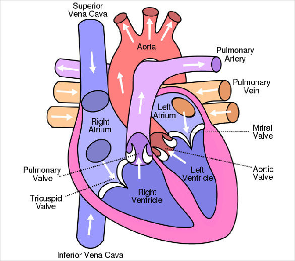

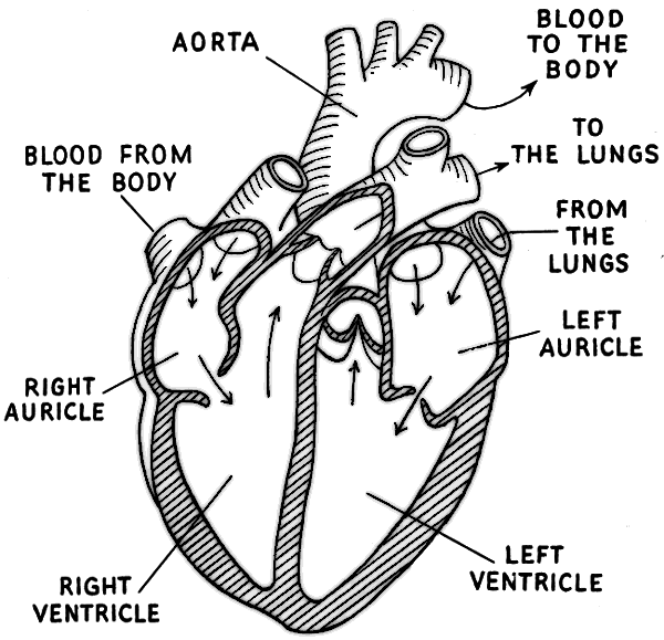


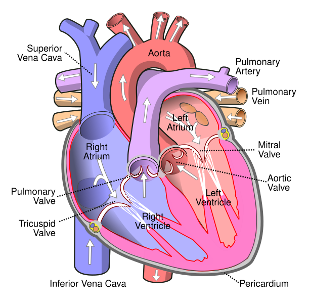






(230).jpg)






Komentar
Posting Komentar